Table of Contents >> Show >> Hide
- What Is fMRI?
- How Does fMRI Work?
- What Is fMRI Used For?
- How Long Does an fMRI Take?
- How to Prepare for an fMRI
- What to Expect During an fMRI
- What Does an fMRI Feel Like?
- What Happens After an fMRI?
- Benefits and Limitations of fMRI
- Final Thoughts
- Experiences Related to fMRI: What People Commonly Notice Before, During, and After the Scan
If standard MRI is the brain’s portrait photographer, fMRI is the nosy documentary crew following it around asking, “Okay, but what are you doing right now?” Functional magnetic resonance imaging, better known as fMRI, is a specialized type of MRI that helps doctors and researchers see which parts of the brain become more active during certain tasks or while the brain is at rest. It does not read your thoughts, reveal whether you secretly dislike kale, or turn your head into a glowing sci-fi orb. What it does do is provide a fascinating, noninvasive way to map brain activity by tracking subtle changes in blood flow.
That makes fMRI useful in several important settings. It can help surgeons identify areas involved in speech, movement, memory, or sensation before brain surgery. It can also help specialists study how a brain affected by stroke, tumor, epilepsy, or other neurological conditions is functioning. In research, fMRI has been a star player for studying attention, language, emotion, decision-making, pain, and a long list of other brain functions.
If your doctor has ordered an fMRI, you may be wondering what exactly happens during the scan, how long it takes, whether it hurts, and whether you’ll be expected to solve math problems while trapped in a noisy tube. Fair questions. Here is what fMRI is, how it works, why it is used, how long it usually takes, and what you can realistically expect before, during, and after the test.
What Is fMRI?
fMRI stands for functional magnetic resonance imaging. Like a standard MRI, it uses a powerful magnet, radio waves, and computer processing to create images. The key difference is that a regular MRI mainly shows structure, while fMRI focuses on function. In other words, a regular MRI shows what the brain looks like, while fMRI helps show what parts of the brain are more active during a task or in a resting state.
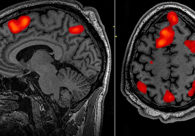
The most common form of fMRI is called BOLD fMRI, which stands for blood oxygen level-dependent imaging. That sounds complicated, but the basic idea is pretty simple: when a region of the brain works harder, it uses more oxygen and usually receives more blood flow. fMRI detects those tiny changes and uses them to build activity maps of the brain.
That is why fMRI is often described as an indirect measure of brain activity. It does not record neurons firing the way an EEG does. Instead, it tracks the changes in blood flow and oxygen use that tend to accompany neural activity. Think of it as seeing the traffic around a stadium and correctly guessing that a concert or game is happening inside.
How Does fMRI Work?
The BOLD signal: the brain’s oxygen clue
Your blood contains hemoglobin, the protein that carries oxygen. Oxygen-rich and oxygen-poor blood behave slightly differently in a magnetic field. When a part of the brain becomes active, the body sends more blood to that area. This changes the local balance between oxygenated and deoxygenated blood. The MRI scanner can detect those tiny shifts, and software turns them into images showing where activity is increasing or decreasing.
That means the colorful fMRI brain maps you have probably seen are not photographs of thoughts lighting up like neon signs. They are statistical maps that represent changes in the BOLD signal over time. Very cool, very useful, and slightly less magical than the internet sometimes makes it sound.
Task-based fMRI
In task-based fMRI, you are asked to do something specific while the scan is running. The task may be physical, verbal, or mental. Common examples include:
- tapping your fingers or moving your hand
- looking at pictures or flashing lights
- listening to words or sounds
- answering simple questions
- doing a language task, such as word generation
- performing a simple mental exercise like basic math
The care team compares brain activity during the task with activity during rest. This helps identify which brain regions are involved in specific functions, such as language, movement, or memory.
Resting-state fMRI
Not every fMRI requires active tasks. In resting-state fMRI, you may simply lie still with your eyes open or closed while the scanner measures spontaneous patterns of brain activity. This can help show how different brain networks communicate when you are not doing a structured task. Researchers love this approach, and clinicians may use it in selected cases, especially when a person cannot easily perform task-based testing.
Why staying still matters so much
fMRI is incredibly sensitive to movement. Even small head movements can blur the data or make the results harder to interpret. So yes, the old MRI instruction to “please hold still” becomes even more important here. During an fMRI, being a statue is basically a job requirement.
What Is fMRI Used For?
fMRI has both clinical and research uses. In everyday patient care, one of the most important uses is presurgical brain mapping. Before brain surgery or certain types of radiation treatment, doctors may need to know exactly where functions like speech, hand movement, or memory are located in that specific person’s brain. Brain anatomy is similar across people, but function is not identical from one person to another, so personalized mapping matters.
Common clinical uses may include:
- Planning brain surgery: especially for tumors, seizure-related surgery, or lesions near areas controlling speech, movement, or sensation.
- Supporting treatment planning: including some cases involving radiation therapy or other invasive brain treatments.
- Evaluating functional effects of disease: such as stroke, brain tumors, traumatic brain injury, or neurological disorders.
- Studying recovery and brain reorganization: for example, how the brain adapts after injury.
In research, fMRI is used to study how the brain handles attention, emotion, language, memory, reading, pain, problem-solving, decision-making, and social behavior. It has also been used in studies of depression, anxiety, Alzheimer’s disease, autism, chronic pain, addiction, and many other conditions. That said, it is important to remember that not every research use is a routine clinical use. In the United States, the best-established approved clinical role is surgical planning and brain mapping.
How Long Does an fMRI Take?
The answer most people want is: usually about an hour, though it can vary.
The actual fMRI scan itself is often completed within about 45 to 60 minutes. Some scans are shorter, and some take longer if your doctor also wants standard MRI images, contrast-enhanced images, or extra sequences to collect more information. Your total time in the imaging center may be longer because of check-in, safety screening, changing clothes, setup, and post-scan review.
So if you are trying to plan your day, “quick coffee break” is probably too optimistic. “A decent-sized appointment” is more accurate.
How to Prepare for an fMRI
Preparation is usually straightforward, but details can vary by facility. Your imaging center may give you specific instructions based on the reason for the scan, whether contrast might be used, and whether you have any medical implants or special needs.
Before your appointment
- Tell your care team about any implanted device, metal in your body, or previous surgeries.
- Mention pacemakers, aneurysm clips, cochlear implants, stimulators, insulin pumps, glucose monitors, stents, filters, or metal fragments.
- Tell them if you are pregnant or might be pregnant.
- Let them know if you have claustrophobia, chronic pain, or trouble lying flat.
- Ask whether you should take your regular medicines as usual. In many cases, the answer is yes, but always follow the instructions you are given.
- Wear comfortable clothes or be prepared to change into a gown.
- Leave jewelry, watches, piercings, and other metal items at home when possible.
Some centers also do a quick language or motor screening before the exam so they can tailor the tasks to you. This is especially helpful when the goal is presurgical mapping.
What about eating, drinking, and caffeine?
This is one of those “follow your facility’s instructions” situations. Some MRI exams have no restrictions, while others may have specific instructions. For fMRI in particular, centers may want your routine to be as typical as possible because caffeine and other factors can influence physiology. If you receive written instructions, follow those instead of guessing.
What to Expect During an fMRI
When you arrive, you will complete a detailed safety questionnaire. This is not paperwork theater. MRI magnets are powerful, and screening is essential. After that, you will remove metal objects and change clothes if needed.
You will lie on a narrow table that slides into the scanner. For brain fMRI, your head usually rests in a special device called a head coil. It is there to improve image quality, not to audition you for a robot movie. You will usually get earplugs or headphones because the scanner can be very loud, making banging, knocking, and humming sounds during image collection.
Once the scan starts, the technologist monitors you from another room, but you are not abandoned to the magnetic void. You can communicate through an intercom, and many centers give you a squeeze bulb or call button if you need help.
During the tasks
If you are having task-based fMRI, the team will guide you through simple activities. You may be asked to:
- tap your thumb to your fingers
- move your hand, foot, or tongue
- name words in your head
- read or listen to language prompts
- answer easy questions
- perform basic mental exercises
Tasks are often done in short blocks. For example, you may do a task for several seconds, then rest, then repeat. The pattern helps the scanner compare activity and identify the corresponding brain regions.
Will contrast be used?
Not always. Some fMRI exams are done without contrast, while others may include standard MRI sequences with gadolinium contrast if the doctor needs additional structural detail. If contrast is planned, the staff will explain why and screen for kidney issues or previous reactions.
What Does an fMRI Feel Like?
For most people, an fMRI is painless. The hardest part is usually not pain but patience. You may notice:
- loud tapping or thumping noises
- a snug, enclosed space
- the need to stay very still
- a slight sense of warmth in the area being scanned
- mental fatigue if the tasks require concentration
If you are prone to claustrophobia, say so before the scan. Facilities may offer strategies such as coaching, extra communication, or medication in some situations. However, because many fMRI exams require you to stay alert and follow instructions, sedation is not always ideal. This is another reason it is smart to bring up anxiety early rather than heroically suffering in silence.
What Happens After an fMRI?
Most people can go home shortly after the scan. If you did not receive a sedating medication, there is usually no recovery period and you can typically resume normal eating, activity, and routine tasks. If you were given medication to relax, you may need someone else to drive you home.
The images are then reviewed and analyzed by specialists, often including a radiologist and, depending on the case, a neurologist, neurosurgeon, or neuropsychologist. Results are not always immediate because fMRI interpretation can be more complex than simply glancing at a picture and saying, “Yep, that is definitely a brain.” The final report is usually sent to the doctor who ordered the scan, who will discuss what it means in the context of your care.
Benefits and Limitations of fMRI
Benefits
- It is noninvasive.
- It does not use ionizing radiation.
- It can show both brain structure and function when combined with standard MRI.
- It helps with personalized brain mapping before treatment.
- It has transformed neuroscience research.
Limitations
- It measures blood-flow changes, not neurons directly.
- It is sensitive to movement, which can affect accuracy.
- Not every person can safely undergo MRI because of certain implants or metal objects.
- Some people find the scanner uncomfortable or claustrophobic.
- Research findings do not always translate into routine clinical decisions.
In short, fMRI is powerful, but it is not magic. It is one tool among many, and it works best when interpreted by experienced professionals alongside other imaging, exams, and clinical information.
Final Thoughts
Functional MRI gives doctors and researchers a remarkable window into the working brain. By tracking small changes in blood flow, it can help map language, movement, sensation, memory, and other functions in ways that standard structural imaging alone cannot. For patients, it is most often useful when precision matters, especially before brain surgery or other treatments near important functional areas.
If you have an fMRI scheduled, the headline is reassuring: the test is usually painless, does not use radiation, and is commonly completed in about an hour. The biggest challenges are the noise, the stillness, and the mental patience required to follow instructions while your brain quietly stars in its own data-driven documentary. Glamorous? Not exactly. Helpful? Absolutely.
Experiences Related to fMRI: What People Commonly Notice Before, During, and After the Scan
One of the most common experiences people report before an fMRI is a mix of curiosity and nerves. Many have already heard of MRI, but the word functional makes the test sound more intense than it usually is. Some imagine a machine that can somehow “watch thoughts happen” in real time. Others worry they will mess up the tasks, move too much, or feel trapped in the scanner. In reality, most people find that the hardest part is not the task itself but simply the anticipation. Once the technologist explains what will happen, the process often feels much more manageable.
During the scan, people frequently say the noise is the biggest surprise. Even when they are warned in advance, the banging and thumping can still be startling the first time. The second most common comment is that staying still takes more effort than expected. It is not painful, but lying in one position while focusing on instructions can make time feel a little stretchy. A five-minute sequence can feel like either thirty seconds or half a semester, depending on your stress level.
Task-based fMRI creates its own unique experience. Some people find the tasks oddly entertaining. Finger tapping, word generation, picture viewing, and simple mental exercises can make the exam feel more interactive than a standard MRI. Others say the tasks become strangely difficult because the scanner is loud and the setting is unfamiliar. A person who can do simple math perfectly well at the kitchen table may suddenly feel as if basic subtraction has become a competitive sport. That does not mean anything is wrong. It usually just means you are in a noisy tube trying not to move your head while following instructions.
People who have claustrophobia often describe the beginning of the exam as the most challenging part. Once they realize they can communicate with the staff, hear directions, and know the scan is moving along in steps, many settle down. The sense of control matters. Knowing there is an intercom, a call device, and a real human nearby makes a big difference. Ear protection also helps more than people expect, not just because it softens the sound, but because it makes the whole environment feel less chaotic.
After the scan, many people are surprised by how ordinary they feel. There is usually no dramatic post-test moment. If no sedative was used, most can get up, leave, and return to normal activities pretty quickly. The emotional reaction afterward is often simple relief. The unknown has become known. The giant scanner is no longer a mysterious machine from a medical thriller but just a very expensive, very noisy tool that helped collect useful information. For patients preparing for surgery or further treatment, that information can be incredibly meaningful, because it helps the care team plan with greater precision and confidence.