Table of Contents >> Show >> Hide
- What Is the Right Atrium? (Definition in Plain English)
- Right Atrium Function: The “Three Jobs” That Keep Blood Moving
- Right Atrium Anatomy: A Guided Tour (No Flashcards Required)
- The Right Atrium’s Secret Side Hustle: Running the Heartbeat
- How the Right Atrium Works During a Normal Heartbeat
- Why Right Atrium Health Matters (Even If You’ve Never Thought About It Once)
- How Clinicians Evaluate the Right Atrium
- Keeping the Right Atrium in Your “Good Books”
- Conclusion
- Experiences Related to Right Atrium Function, Definition & Anatomy (Real-World Moments)
If the heart were a busy airport, the right atrium would be arrivals: it’s where “used” (oxygen-poor)
blood checks back in before getting sent off to the lungs for a fresh oxygen makeover. It’s also where the heartbeat’s
main timekeeper livesso yes, this chamber is both the front desk and the drummer in the band.
In this guide, we’ll break down what the right atrium is, what it does minute-to-minute, what it looks like inside,
and why doctors care about it in real life (hint: it’s not just because anatomy exams enjoy causing stress).
What Is the Right Atrium? (Definition in Plain English)
The right atrium (RA) is one of the heart’s four chambers and is located in the upper-right portion of the heart.
It’s an intake chamber: it receives oxygen-poor blood returning from the body and funnels it toward the right ventricle,
which then pumps it to the lungs.
Think of the right atrium as a flexible holding room. It doesn’t need to generate huge pressure like a ventricle.
Its superpower is timing, stretch, and flow managementso the right ventricle gets filled
efficiently without chaos.
Right Atrium Function: The “Three Jobs” That Keep Blood Moving
Modern cardiology often describes atrial performance in three phases. The right atrium is not just a passive cup; it’s a
dynamic chamber that changes roles during the cardiac cycle.
1) Reservoir: the “Storage Mode”
When the tricuspid valve is closed and the right ventricle is contracting, blood continues returning from the body. The right atrium
acts as a reservoir, stretching to store that blood briefly.
2) Conduit: the “Open-Hallway Mode”
When the tricuspid valve opens, the right atrium becomes a conduit. Blood flows from the veins through the right atrium
and into the right ventricle with minimal resistancelike opening the doors so the crowd can move straight through.
3) Booster Pump: the “Final Nudge”
Near the end of ventricular filling, the right atrium contracts (atrial systole) to give the right ventricle an extra
top-off. This “atrial kick” can matter more when the ventricle is stiff or when the heart is working harder.
Bottom line: the right atrium helps optimize right-ventricular filling, supports steady cardiac output, and adapts to
changes in breathing, posture, exercise, and fluid volume.
Right Atrium Anatomy: A Guided Tour (No Flashcards Required)
The right atrium has a few key internal landmarks that explain both its function and its involvement in rhythm problems.
If you ever wondered why anatomy people get weirdly excited about ridges and pits, this is their moment.
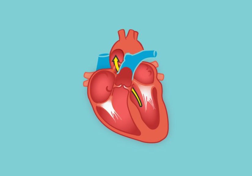
Where Blood Enters the Right Atrium
- Superior vena cava (SVC): brings oxygen-poor blood from the upper body (head, arms, upper chest).
- Inferior vena cava (IVC): returns oxygen-poor blood from the lower body (abdomen, pelvis, legs).
- Coronary sinus: drains blood from the heart muscle itself back into the right atrium.
Where Blood Leaves the Right Atrium
Blood exits the right atrium through the tricuspid valve into the right ventricle. The valve is one-way:
it opens for filling and closes to prevent backflow when the ventricle contracts.
Two “Textures”: Smooth Wall vs. Ridged Wall
The right atrium has two visually distinct regions:
- Sinus venarum (smooth portion): a smoother area that receives the vena cavae and coronary sinusbuilt for streamlined flow.
-
Right atrial appendage (trabeculated portion): a more muscular, ridged extension with comb-like ridges called
pectinate muscles.
Crista Terminalis: the “Border Wall” Inside the Right Atrium
Separating the smooth and ridged regions is a muscular ridge called the crista terminalis.
It’s more than anatomical decoration: it marks developmental boundaries and is an important neighborhood for the heart’s natural pacemaker.
Fossa Ovalis: Evidence You Were Once a Fetus
On the interatrial septum (the wall between the atria), the right atrium contains a depression called the fossa ovalis.
It’s the remnant of the fetal foramen ovale, a flap-like pathway that normally closes after birth. If it doesn’t seal completely,
it can remain as a patent foramen ovale (PFO) in some people.
Small “Doorman” Structures: Eustachian Valve & Thebesian Valve
Some hearts have small valve-like folds:
- Eustachian valve: a remnant near the IVC opening (more relevant in fetal circulation than adult life).
- Thebesian valve: a small fold near the coronary sinus opening in some individuals.
These are usually harmless, but they can matter in imaging or certain procedures (because the heart loves to be “interesting” during catheter navigation).
The Right Atrium’s Secret Side Hustle: Running the Heartbeat
The right atrium isn’t only about blood flowit’s also a major player in electrical conduction.
The heart’s natural pacemaker, the sinoatrial (SA) node, is located in the upper right atrium region near the junction with the SVC.
The SA node fires, the atria contract, and the electrical signal ultimately coordinates ventricular contraction.
This is why right-atrial anatomy shows up in discussions of arrhythmias (like atrial flutter or atrial fibrillation):
structure and electricity are roommates in the atria.
How the Right Atrium Works During a Normal Heartbeat
Let’s follow a single heartbeat in “movie trailer” form:
- Venous return: Blood flows in from the SVC, IVC, and coronary sinus into the right atrium.
- Tricuspid valve opens: Blood moves into the right ventricle (conduit phase).
- Atrial contraction: The right atrium contracts to push the last bit of blood into the ventricle (booster phase).
- Ventricle contracts: The tricuspid valve closes, and the right atrium becomes a reservoir again while the ventricle sends blood to the lungs.
A Practical Example: What Breathing Does to the Right Atrium
During inhalation, pressure in the chest tends to drop, which can increase venous return to the right atrium.
That means the right atrium constantly adjustssubtly changing its filling and emptying patterns so the right ventricle
stays appropriately supplied.
Why Right Atrium Health Matters (Even If You’ve Never Thought About It Once)
Right Atrial Enlargement: a “Clue” Chamber
The right atrium may enlarge when it faces chronically increased volume or pressureoften as a downstream effect of
lung disease, pulmonary hypertension, tricuspid valve problems, or congenital shunts.
Enlargement is not a diagnosis by itself; it’s a sign that invites a bigger investigation.
Arrhythmias: When the Rhythm Neighborhood Gets Noisy
Because important conduction pathways are associated with the right atrium, changes in atrial size, pressure, or tissue structure
can be associated with rhythm issues. A classic example is atrial flutter, which frequently involves circuits in the right atrium.
Atrial fibrillation also involves atrial remodeling and altered atrial function.
ASD, PFO, and the Interatrial Septum
Openings or defects in the septum between the atria can alter blood flow patterns. Some defects are present from birth,
while a PFO is a persistently open flap that is common and often silent. In some scenarios, these openings can be clinically meaningful,
especially if they allow abnormal shunting of blood between chambers.
Fluid Overload & Hormone Signaling (Yes, Really)
The right atrial appendage can release natriuretic peptides when stretched by increased volume.
In everyday terms: when the chamber senses “too much fluid,” chemical signals can help the body reduce volume by
widening blood vessels and increasing salt-and-water excretion through urine.
How Clinicians Evaluate the Right Atrium
Echocardiogram (Ultrasound)
Echocardiography is commonly used to estimate right atrial size, assess right-sided filling, evaluate tricuspid valve function,
and look for signs of elevated right-sided pressures or shunts. Advanced echo methods can also analyze atrial strain (how the tissue deforms as it fills and empties).
ECG (Electrocardiogram)
ECG patterns can sometimes suggest right atrial enlargement or atrial rhythm disturbances. The ECG doesn’t “photograph” the chamber,
but it can provide electrical fingerprints that prompt further evaluation.
CT or MRI
Cross-sectional imaging (CT or cardiac MRI) can provide detailed structural informationuseful when anatomy is complex
or when physicians need high-resolution views for procedural planning or congenital heart assessment.
Right Heart Catheterization
When clinicians need direct measurements of pressures in the right side of the heart and lungs, a right heart catheterization
may be used. This test can help assess how effectively the heart is pumping and quantify pressures that reflect pulmonary circulation and right-heart loading.
Keeping the Right Atrium in Your “Good Books”
You can’t directly “work out” your right atrium like a bicep, but you can support right-heart health by supporting the systems that affect it:
lung health, blood pressure, fluid balance, and heart rhythm stability.
- Protect lung function: Avoid smoking/vaping, treat chronic lung disease, and follow medical guidance for asthma/COPD.
- Manage blood pressure: High pressures in the pulmonary circuit or left-heart strain can indirectly burden the right side.
- Stay active within your limits: Aerobic conditioning supports overall cardiovascular efficiency.
- Take symptoms seriously: New palpitations, fainting, unusual shortness of breath, or swelling deserve medical evaluation.
If you have known heart or lung disease, individualized guidance from a clinician is essentialbecause the right atrium responds to the
whole-body system, not just a single isolated “heart part.”
Conclusion
The right atrium is the heart’s upper-right gateway: it receives oxygen-poor blood from the body and heart muscle, coordinates smooth flow through the
tricuspid valve into the right ventricle, and plays a key role in heart rhythm through its relationship to the SA node.
Its anatomysinus venarum, pectinate muscles, crista terminalis, and the fossa ovalisexplains why it’s involved in everything from
normal filling mechanics to arrhythmias and congenital septal issues.
In other words: the right atrium may not get the glamorous “big pump” reputation, but it’s doing quiet, essential work every second you’re alive.
(And it does it without asking for a standing ovation, which is honestly very mature of it.)
Experiences Related to Right Atrium Function, Definition & Anatomy (Real-World Moments)
You don’t “feel” your right atrium working the way you feel a muscle burn during a workout. Most of the time, it’s a silent teammate.
But the right atrium shows up in real life in a few surprisingly relatable waysthrough symptoms people notice, through what clinicians see on tests,
and through the lived experience of learning how the heart actually behaves in a moving, breathing human.
1) The “Why Am I Out of Breath?” Experience
A common pathway to right-atrial conversations starts with a simple complaint: “I’m more winded than I used to be.”
When lungs or pulmonary blood vessels are under strain (for example, from pulmonary hypertension or chronic lung disease),
the right side of the heart works harder. People may describe shortness of breath on stairs, reduced exercise tolerance,
or fatigue that feels out of proportion to effort. Clinicians then look at the right heartoften starting with an echocardiogramto see whether
the right atrium is enlarged or whether right-sided pressures appear elevated.
2) Palpitations: When the Rhythm Gets Your Attention
Another common lived experience is the sudden awareness of the heartbeat itself: fluttering, pounding, racing, or a “fish flopping” sensation in the chest.
Because the right atrium is intimately tied to electrical activation (and right-atrial tissue can participate in arrhythmia circuits),
rhythm problems can turn the right atrium from background character to lead actor. People often report that palpitations are worse with stress,
poor sleep, dehydration, alcohol, or stimulantsfactors that can tip a sensitive electrical system into louder behavior.
While many palpitations are benign, the experience is unsettling, and it’s one reason clinicians pair symptom descriptions with ECGs and sometimes monitors.
3) The Catheter Lab “Tour” (From the Inside, Sort Of)
For patients who undergo right heart catheterization, the right atrium becomes more than a diagram. They may remember hearing terms like
“right atrial pressure” and “pulmonary artery pressure,” and feeling reassured (or anxious) as numbers are interpreted in real time.
Many people describe the experience as less dramatic than expectedmore like a carefully choreographed procedure with lots of calm voices,
sterile drapes, and steady monitoring. The right atrium is literally part of the route a catheter takes to measure pressures and evaluate how the
heart and lungs are working together.
4) Learning Anatomy: From Flat Pictures to 3D Reality
Students often have a “click” moment with the right atrium when they realize the inside isn’t a smooth bowl. The pectinate muscles, the crista terminalis,
and the fossa ovalis turn the chamber into a textured landscape with landmarks that matter. The “aha” is usually this:
anatomy isn’t triviait’s a map for function and procedures. The crista terminalis is not just a ridge; it helps define regions and relates to where the
SA node lives. The fossa ovalis isn’t just a dent; it’s a clue to fetal circulation and the possibility of a PFO. These connections make the right atrium
feel less like a label and more like a working space with history and purpose.
5) A Simple Everyday Example: The “Big Meal + Tight Pants” Phenomenon
Here’s a surprisingly human experience: some people notice stronger heartbeats or mild breathlessness after a very large meal, especially if they’re
also sedentary or wearing restrictive clothing. No, your right atrium isn’t sending you a passive-aggressive text. But circulation, diaphragm motion,
and venous return can shift with posture and abdominal pressure. The right atrium is one of the first heart chambers to deal with those venous-return changes,
adjusting its filling behavior as your body redistributes blood. It’s a small reminder that heart chambers don’t work in isolationthey respond to the
whole-body context you’re living in.
The takeaway from these experiences is simple: the right atrium is a master adapter. Most of the time, it’s invisible.
But when breathing, volume status, or rhythm gets disrupted, it becomes a meaningful window into what’s happening in the cardio-pulmonary system.