Table of Contents >> Show >> Hide
- What Is a Heart PET Scan?
- Purpose: Why Would Someone Need a Heart PET Scan?
- How to Prepare for a Heart PET Scan
- Procedure: What Happens During the Scan?
- How Doctors Read the Results (in Plain English)
- Risks: What Can Go Wrong (and How Often It’s Serious)
- Questions Worth Asking Before Your Scan
- Bottom Line
- Patient Experiences: What It Often Feels Like (Added ~)
- SEO Tags
If your doctor ordered a heart PET scan, you might be imagining a futuristic machine reading your thoughts, or at least your feelings about
treadmill running. (Good news: it does neither.) A cardiac PET scan is a highly detailed imaging test that helps your care team understand how well blood is
reaching your heart muscleand, in some cases, whether parts of your heart are “asleep but salvageable” or actively inflamed.
This guide pulls together how heart PET scans are typically explained by major U.S. medical centers and cardiology/radiology organizations (think:
large hospital systems, nuclear cardiology societies, and patient education resources). We’ll cover what the test is for, what happens step-by-step, how to
prepare, what the results can mean, and the real-world riskswithout turning it into a textbook or a horror movie.
What Is a Heart PET Scan?
A heart PET scan (also called cardiac PET) is a nuclear medicine test that uses a tiny amount of a radioactive “tracer” plus
a special scanner to create images of your heart. The tracer travels through your blood, and the scanner detects the signal it gives off. That signal is used
to build detailed pictures showing how well blood is flowing to different areas of the heart muscle.
Many heart PET scans are done as PET myocardial perfusion imaging (PET MPI). “Perfusion” is just a fancy way of saying “blood flow.”
PET MPI is often paired with a stress testeither exercise (treadmill) or medication that makes the heart work harderso doctors can compare
blood flow at rest versus stress.
PET vs. other heart tests (why PET is a big deal)
- Sharper imaging: PET tends to produce clearer images and can reduce certain “false alarms” caused by soft tissue or body shape.
-
Quantitative blood flow: Many PET systems can measure how much blood is moving through the heart muscle and calculate
myocardial flow reserve (a key clue for microvascular disease). - Often faster: Depending on the tracer and protocol, PET MPI can be quicker than some older nuclear imaging approaches.
Purpose: Why Would Someone Need a Heart PET Scan?
Your doctor doesn’t order a heart PET scan just because the machine looks cool (it does) or because your heart deserves a photoshoot (it does, but still).
Typically, it’s ordered when the care team needs a high-confidence answer about blood flow, risk, or heart muscle healthespecially when other tests are
inconclusive or when more detail matters.
1) Check for coronary artery disease (CAD)
The most common reason is to look for coronary artery diseasenarrowing or blockage in the arteries that supply the heart. If a section of
the heart muscle gets less blood during stress than at rest, that can suggest a significant narrowing that might explain symptoms like chest pressure,
shortness of breath, or unusual fatigue.
2) Evaluate chest pain when the picture is complicated
PET can be especially helpful when standard tests are harder to interpretlike in people with a higher body weight, breast tissue that can affect imaging
angles, or prior heart procedures. The goal is to reduce “maybe” and get closer to “here’s what’s happening.”
3) Measure coronary flow reserve and detect microvascular dysfunction
Not all heart-related chest pain comes from big artery blockages. Some people have symptoms because the tiny vessels in the heart don’t
widen properly under stress (often called coronary microvascular dysfunction). PET can quantify blood flow and calculate
myocardial flow reserve, which can help identify this problem even when major arteries look “not too bad” on other tests.
4) See if heart muscle is viable (alive) after damage
In people with heart failure or prior heart attacks, doctors may use PET to assess viabilitywhether weakened areas of the heart muscle are
scarred beyond recovery or still alive but underfed. This can matter when deciding whether procedures like bypass surgery or certain interventions are likely
to improve function.
5) Look for inflammation or infection in specific situations
Some cardiac PET scans use a glucose-based tracer (often called FDG PET) to look for areas of active inflammation. This can be used in
selected cases like suspected cardiac sarcoidosis or certain infection evaluations, usually in specialized centers with specific prep
protocols.
How to Prepare for a Heart PET Scan
Preparation depends on which type of cardiac PET you’re having (perfusion vs. inflammation/FDG protocols), and your clinic will give exact
instructions. But there are common themesespecially around food, caffeine, and medications.
Common prep for PET myocardial perfusion (blood flow) studies
- Fasting: Many sites ask you to avoid food for several hours before the test (water is usually okay).
-
No caffeine for about 24 hours: Coffee, tea, energy drinks, soda, chocolate, and even “decaf” can interfere with medication-based stress
testing and the results. -
Medication adjustments: Some heart meds (and asthma meds) may need special handling before a stress study. Never stop a medication on your
ownfollow the instruction sheet or call the clinic. -
Wear comfy clothing: You’ll be lying still, and sometimes you’ll walk on a treadmill. Dress like you’re meeting a machine that loves
sweatpants.
Prep for FDG PET inflammation protocols (like cardiac sarcoidosis)
FDG PET often requires more specific preparation because the goal is to reduce normal glucose uptake in the heart muscle so inflammatory “hot spots”
stand out clearly. Many protocols use a high-fat, very-low-carb approach the day before and a prolonged fast, though exact instructions vary.
Pro tip: preparation is part of the test
Cardiac PET is powerful, but it’s not magic. Caffeine, certain medications, and eating patterns can change the physiology the scan is trying to measure.
If the prep feels picky, it’s because your doctors want the images to answer the question accurately the first time.
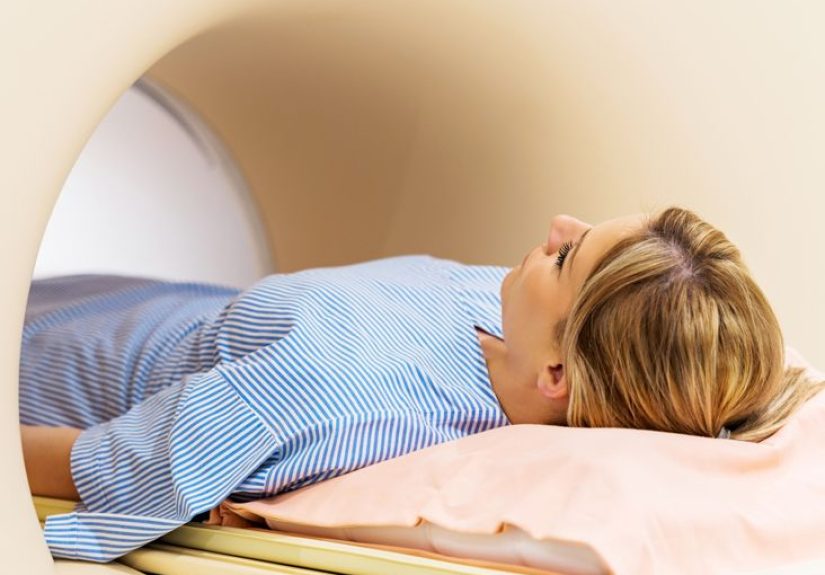
Procedure: What Happens During the Scan?
A heart PET scan is usually an outpatient test. You’ll check in, answer safety questions, get an IV, and spend part of the visit lying still while the scanner
does its job. Here’s what a typical PET MPI (perfusion) visit often looks like.
Step 1: Check-in and safety screening
Staff will confirm your medications, allergies, and whether there’s any chance of pregnancy. They’ll also ask about asthma/COPD symptoms, because some stress
medications can be a problem for certain lung conditions.
Step 2: IV placement
A technologist places an IV line, usually in your arm. The tracer and (if needed) stress medication are delivered through this IV. The most common “drama” at
this stage is mild pinchingnothing like movie-level needle scenes.
Step 3: Rest imaging
You’ll receive a small dose of radiotracer. After a short waiting period (so it can circulate), you’ll lie on the scanning table. The scanner is shaped like a
large donut. It does not touch you, but it does appreciate when you stay still.
Step 4: Stress portion (exercise or medication)
Next comes “stress.” Some people walk on a treadmill. Many cardiac PET perfusion tests use medication to widen coronary arteries and simulate exercise-like
conditions. Common agents include regadenoson or adenosine-type drugs; some protocols use dobutamine in specific cases.
During this portion, your heart rhythm, blood pressure, and symptoms are monitored closely. You may feel flushing, shortness of breath, chest pressure, or a
“weird” sensation that’s hard to describe. The good news: these feelings are usually brief, and staff can treat side effects quickly.
Step 5: Stress imaging
A second tracer dose is given during peak stress (or right after exercise), followed by another set of images. Many PET systems also perform a low-dose CT scan
for attenuation correction, which helps refine image accuracy.
Step 6: Wrap-up
After imaging, you’ll usually be encouraged to drink fluids to help your body clear the tracer. Most people can return to normal activities the same day unless
their clinic gives specific restrictions.
How Doctors Read the Results (in Plain English)
Your cardiologist and imaging team look at:
-
Perfusion patterns: Are there areas that get less blood during stress than at rest? That “stress-only dip” can suggest a flow-limiting
narrowing. - Extent and severity: Is the problem small and mild, or large and intense?
- Heart function: Many studies also estimate how well the heart pumps (ejection fraction) and how it moves.
- Blood flow quantification: If measured, myocardial blood flow and myocardial flow reserve help assess diffuse disease or microvascular issues.
Example: Suppose your scan shows a reversible perfusion defect in a region supplied by a specific coronary artery. That finding may steer your doctor toward
adjusting medications, ordering a CT angiogram or catheterization, or considering revascularizationdepending on symptoms, overall risk, and how much heart muscle
is affected.
Another example: If major arteries don’t show a focal defect, but your flow reserve is reduced, your doctor may talk about microvascular dysfunction and focus
on targeted medical therapy and risk-factor control rather than procedures meant for a single blockage.
Risks: What Can Go Wrong (and How Often It’s Serious)
The overall risk of a heart PET scan is usually low, but “low” doesn’t mean “zero.” It’s more like: the test is designed to be safe, monitored, and worth it
when it’s ordered for the right reason.
Radiation exposure
PET uses a small amount of radiation from the tracer (and sometimes a low-dose CT component). For most people, the risk from this level of exposure is
considered very small compared with the benefit of accurate diagnosis. However, pregnancy and breastfeeding require special discussion because radiation can
affect a fetus or infant.
IV-related issues
- Bruising or soreness where the IV was placed
- Rare tracer leakage into surrounding tissue (usually uncomfortable, typically not dangerous)
- Allergic reactions to radiotracers are uncommon and usually mild, but they can happen
Stress-test medication side effects
Medication stress agents can cause temporary symptoms such as flushing, headache, nausea, shortness of breath, dizziness, or chest discomfort. Serious events
(like dangerous rhythm problems or heart attack) are rare, but they’ve been reportedthis is why you’re monitored during the test and why clinics ask detailed
questions about heart and lung history beforehand.
False positives/false negatives (the “test interpretation” risk)
Not every abnormal scan means a major blockage, and not every normal scan guarantees “nothing is going on.” Preparation issues (especially caffeine), movement
during imaging, or certain medical conditions can blur interpretation. That’s also why doctors interpret results in contextsymptoms, history, labs, and other
imaging.
Questions Worth Asking Before Your Scan
- Is this a perfusion PET (blood flow) study, an FDG inflammation study, or both?
- Exactly when do I need to stop caffeine, food, and certain medications?
- If I have asthma/COPD, what stress agent will you use and why?
- How long will I be at the clinic from check-in to check-out?
- How and when will I get results, and what are the likely next steps based on different outcomes?
Bottom Line
A heart PET scan is one of the most informative noninvasive tools for understanding heart blood flowand, in select cases, heart muscle viability
or inflammation. It’s precise, monitored, and designed to answer practical clinical questions: “Is there a problem?” “How big is it?” and “What should we do
next?”
If you’re feeling nervous, that’s normal. But most people finish the test thinking, “Okay… that was more awkward than scary.” Follow the prep instructions,
tell the team what you’re feeling during the stress portion, and remember: the whole point is to get a clearer map so your care plan can be smarter.
Patient Experiences: What It Often Feels Like (Added ~)
People tend to show up to a cardiac PET scan with three questions: “Will it hurt?” “Will I feel trapped?” and “How weird is the stress medication going to
be?” The most common surprise is that the test is more like a carefully choreographed routine than an intense medical ordeal. You check in, answer a lot of
questions (yes, they really do want to know about caffeine), and then the pace slows down once you’re in the imaging area.
The IV is often the most uncomfortable part for many patientsmostly because it’s a pinch plus a little pressure. After that, the rest portion
can feel almost boring in a good way. You lie still, the scanner hums, and the technologist may remind you not to move. Some people describe the scanner as
“close but not claustrophobic,” because it’s open like a ring rather than a long tube. If you’re someone who hates staying still, this is the moment when your
brain suddenly invents ten urgent reasons to scratch your nose. (Try to resist. The images will thank you.)
The stress portion is where experiences vary the most. If you’re doing treadmill exercise, people often say it feels like a regular stress
testonly with more staff attention and a stronger sense that your heart is the main character today. If you’re getting a medication stress agent, patients
commonly describe a fast-onset wave of sensations: warmth, flushing, heavier breathing, sometimes mild chest pressure, and a “this is odd” feeling that’s hard
to label. The key detail is that these sensations usually pass quickly. Many patients feel noticeably better within a minute or two, and clinics can give
medications to reverse lingering effects if needed.
Another common experience is worry about what symptoms “mean.” People may think, “If I feel short of breath, does that mean something is wrong?” Not
necessarilysome stress agents are known to cause that sensation even when the heart is okay. That’s why the team watches your EKG and blood pressure and asks
you to describe what you feel. If you speak up early“I’m flushed,” “I feel tightness,” “I’m dizzy”staff can respond quickly, and you’ll usually feel more in
control of the process.
After the scan, many people report feeling relieved more than anything. You can usually eat, hydrate, and return to normal activities (unless
instructed otherwise). Some notice mild fatigue, which can be from fasting, the stress agent, or just the adrenaline of having a medical test. The emotional
side is real too: waiting for results can be the hardest part. Patients often say it helps to ask upfront when results will be reviewed and whether you’ll hear
from the ordering doctor by phone, portal message, or follow-up visitbecause uncertainty is a sneaky stress test all by itself.
Overall, the most consistent “review” you’ll hear is: the preparation matters, the stress sensations can be strange but brief, and the staff presence
is reassuring. A heart PET scan is less like an ordeal and more like a highly supervised science experimentwith you as the star participant and your heart as
the data set everyone is rooting for.