Table of Contents >> Show >> Hide
- Quick Navigation
- Definition: What Calciphylaxis Is (and Isn’t)
- Calciphylaxis Pictures: What You’ll See (and Why They’re Hard to Google at 2 a.m.)
- Symptoms: How It Feels and How It Progresses
- Who’s at Risk (and Why Kidneys Matter)
- Diagnosis: How Doctors Confirm It
- Treatment: What Helps, What’s Debated
- 1) Stabilize the patient and control pain
- 2) Wound care and infection prevention
- 3) Correct mineral imbalance (calcium/phosphate/PTH)
- 4) Sodium thiosulfate (a common off-label therapy)
- 5) Review medications and remove potential contributors
- 6) Adjuncts: hyperbaric oxygen, negative pressure therapy, grafting
- A realistic mini-scenario (how treatment can look)
- Outlook and Prognosis
- Experiences: What It’s Like in Real Life (The Part People Don’t Put in Textbooks)
- The pain is not “just a skin thing”
- Wound care becomes a routine… and a lifestyle
- Dialysis days can feel like “treatment stacking”
- Food becomes complicatedagain
- Medication changes can be scarybut communication helps
- The emotional side is real (and deserves treatment too)
- What patients often wish they’d known sooner
Important note: This article is educational, not medical advice. Calciphylaxis is a medical emergencyif you suspect it, contact a clinician urgently.
Definition: What Calciphylaxis Is (and Isn’t)
Calciphylaxis (also called calcific uremic arteriolopathy, or CUA) is a rare but serious condition where calcium deposits and clotting changes occur in small blood vesselsespecially those supplying the skin and fatleading to poor blood flow, tissue injury, and sometimes skin necrosis (tissue death). The result can be intensely painful skin lesions that may break down into ulcers and become infected.
A quick myth-buster: despite the name, calciphylaxis is not an allergy and it’s not “anaphylaxis with a sprinkle of calcium.” The “-phylaxis” part is historical, not a warning label for your immune system. It’s primarily a disorder of microvascular injurycalcification, narrowing, and thrombosisoften tied to severe kidney disease and mineral-bone imbalance.
Why it happens (in plain English)
Think of small skin arteries as tiny garden hoses. Calciphylaxis can stiffen and narrow those hoses with calcium deposits and thickened vessel walls. Add micro-clots, and blood can’t reach the tissue well. Starved tissue becomes inflamed, then damaged, then (in severe cases) breaks down into ulcers.
It most commonly affects people with end-stage kidney disease on dialysis, but it can also occur in people without dialysis (sometimes called non-uremic calciphylaxis). Either way, it’s uncommonand that rarity is part of why diagnosis and treatment can be challenging.
Calciphylaxis Pictures: What You’ll See (and Why They’re Hard to Google at 2 a.m.)
If you’re searching for “calciphylaxis pictures,” you’re probably trying to answer one urgent question: Does this look like what I’m seeing? The honest answer is tricky because calciphylaxis can resemble other conditions (cellulitis, vasculitis, diabetic ulcers, cholesterol emboli, and more). Also, many clinical photos are graphic and may be restricted on some platforms.
Early-stage appearance (often before ulcers)
- Pain out of proportion to what you see on the surface.
- Violaceous (purple) patches or blotchy discoloration.
- Retiform purpura (a net-like, mottled purple pattern).
- Tender nodules or plaques under the skinlike sore “lumps” that don’t feel like normal bruises.
Later-stage appearance (ulceration and necrosis)
- Open sores (ulcers) that enlarge or deepen.
- Black eschar (a leathery, black scab-like area) from tissue death.
- Drainage, odor, warmth, swellingpossible signs of infection.
Common locations in pictures
Photos frequently show lesions on areas with more fat tissue: thighs, abdomen, buttocks. Lower legs can be involved too. Lesions may be single or multiple, and they can progress quickly.
“Pictures” aren’t only skin photos
Clinicians may also use:
- Imaging images (like X-rays) that can show vascular calcification in soft tissues.
- Biopsy histology pictures showing calcification in small vessels and surrounding tissue changes.
If you’re taking pictures for your doctor
- Use bright, natural light and take 2–3 angles.
- Include a size reference (coin or ruler) next to the lesion.
- Take one close-up and one wider shot showing the location on the body.
- If the pain is severe or the area looks rapidly worse, don’t wait for the “perfect photo.” Seek care.
Symptoms: How It Feels and How It Progresses
Calciphylaxis is notorious for being extremely painful. Many people describe pain that’s burning, deep, or relentlesssometimes starting before the skin looks dramatically different.
Core symptoms
- Severe pain at the site (sometimes out of proportion to early skin findings).
- Purple or mottled skin discoloration, bruised-looking patches, or net-like purpura.
- Firm lumps or plaques under the skin.
- Ulcers that may form and worsen.
- Black, necrotic areas (eschar) in advanced disease.
Red flags that warrant urgent evaluation
- Rapidly expanding painful skin lesions, especially in someone with dialysis or advanced kidney disease.
- Fever, chills, worsening redness, pus, or foul odor (possible infection).
- New confusion, low blood pressure, or feeling “dangerously sick” (possible sepsis).
Why infection is such a big deal
The skin is your body’s security system. When ulcers form, the “doors” are open. Calciphylaxis ulcers can be difficult to heal and may become infected, and infection can become systemic. This is one reason clinicians take calciphylaxis so seriously and treat it with a team approach.
Who’s at Risk (and Why Kidneys Matter)
Calciphylaxis is most commonly associated with advanced chronic kidney disease and dialysis, but it’s not exclusive to kidney failure. Risk is often linked to a perfect storm of mineral imbalance, vascular injury, and pro-thrombotic (clot-promoting) conditions.
Commonly associated factors
- End-stage kidney disease and dialysis (hemodialysis or peritoneal dialysis).
- CKD-mineral and bone disorder: high phosphate, calcium/phosphate imbalance, and secondary hyperparathyroidism.
- Warfarin exposure (reported as a risk factor in multiple reviews).
- Diabetes, obesity, and low albumin (a marker of poor nutrition/inflammation).
- Some medications and conditions that affect coagulation or mineral metabolism (your clinician will review these case-by-case).
Non-uremic calciphylaxis
Calciphylaxis can occur without end-stage kidney disease. Reported associations include primary hyperparathyroidism, certain autoimmune conditions, malignancy, and medications that influence clotting or calcification pathways. It’s rarer, but it’s one reason clinicians avoid assuming, “No dialysis = no calciphylaxis.”
Diagnosis: How Doctors Confirm It
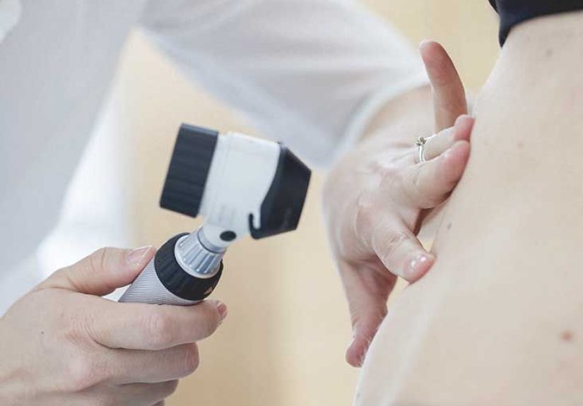
Diagnosis often starts with a high index of suspicion: a person at risk (often dialysis) with severe painful lesions that look like purpura, plaques, or necrotic ulcers. Confirmation can involve labs, imaging, and sometimes biopsybalanced against the fact that skin trauma can worsen wounds.
Clinical exam and history
- Timeline: how fast the lesion appeared and changed.
- Pain intensity and pattern.
- Dialysis history, kidney function stage, recent medication changes (especially anticoagulants and calcium/phosphate-related meds).
Labs that help map the terrain
There’s no single “calciphylaxis blood test,” but clinicians commonly review calcium, phosphate, parathyroid hormone (PTH), vitamin D-related therapy, inflammatory markers, and albuminbecause management usually involves correcting mineral and metabolic issues.
Imaging (sometimes)
Plain radiographs may show soft-tissue or vascular calcification, and some centers use other imaging tools to support diagnosis. Imaging is supportive, not always definitive.
Skin biopsy: helpful, but not always simple
A biopsy can show small-vessel calcification and thrombosis consistent with calciphylaxis. However, biopsy can create a new wound in tissue that already has poor blood flow. Many clinicians weigh biopsy risks versus benefits carefully, and if done, it’s usually performed by experienced teams.
Conditions that can mimic calciphylaxis
- Cellulitis (bacterial skin infection)
- Vasculitis
- Warfarin-related skin necrosis
- Cholesterol embolization
- Diabetic foot/vascular ulcers
- Necrotizing fasciitis (surgical emergency)
In practice, clinicians often treat urgent threats (pain control, infection coverage when indicated) while working toward confirmation. The “don’t miss” diagnosis is the one that gets worse while you’re waiting for perfect certainty.
Treatment: What Helps, What’s Debated
Calciphylaxis treatment is typically multidisciplinary: nephrology, dermatology, wound care, surgery (sometimes), pain management, nutrition, and infectious disease when needed. There is no single universally approved cure, so care is individualized and often combines multiple strategies.
1) Stabilize the patient and control pain
Pain can be severe and may require layered approachestopical measures, systemic medications, and sometimes advanced pain management. No one gets a medal for “toughing it out.” Pain control is part of treatment, not a side quest.
2) Wound care and infection prevention
- Careful cleansing and dressings designed for fragile, ischemic skin.
- Monitoring for infection; antibiotics if infection is suspected or confirmed.
- In some cases, debridement (removing dead tissue) may be consideredespecially when infection or sepsis risk is high.
3) Correct mineral imbalance (calcium/phosphate/PTH)
Because calciphylaxis is linked to abnormal mineral metabolism in many kidney patients, clinicians often work to bring phosphate down, rebalance calcium, and manage secondary hyperparathyroidism. This may include:
- Adjusting phosphate binders (often favoring non-calcium binders when appropriate).
- Dialysis prescription optimization and dietary phosphorus strategies (guided by renal dietitians).
- Cinacalcet to reduce PTH in selected patients, or other therapies depending on the clinical scenario.
- In certain cases, parathyroidectomy may be considered if hyperparathyroidism is severe and refractory.
4) Sodium thiosulfate (a common off-label therapy)
Sodium thiosulfate is frequently used (often intravenously) in dialysis-associated calciphylaxis. Many centers administer it during or after dialysis sessions several times per week. It’s thought to help by influencing calcification chemistry and improving symptoms in some patients, though responses vary and side effects (like nausea and metabolic issues) can occur.
5) Review medications and remove potential contributors
Clinicians often reconsider medications that may worsen riskespecially those affecting clotting or mineral balance. For example, warfarin has been associated as a risk factor in multiple reviews, so teams may evaluate alternatives if anticoagulation is needed. (Do not stop anticoagulants on your own; abrupt changes can be dangerous.)
6) Adjuncts: hyperbaric oxygen, negative pressure therapy, grafting
Depending on the wound and the patient’s overall stability, clinicians may consider adjunctive therapies such as hyperbaric oxygen therapy or negative pressure wound therapy. In selected cases, once a healthy granulation bed forms, skin grafting can be part of reconstruction.
A realistic mini-scenario (how treatment can look)
Imagine a dialysis patientlet’s call her Mariawho develops a painful purple patch on her thigh that rapidly worsens. Her care team moves fast: pain control, labs to review phosphate and PTH, wound care consult, and a discussion of whether a biopsy is worth the risk. Because infection is a major threat, they watch closely for fever, drainage, and rising inflammatory markers. They adjust phosphate binders, consider sodium thiosulfate during dialysis, and review whether any meds (like warfarin) should be changed. The “big win” is early recognitionbefore the wound becomes a deep ulcer.
Outlook and Prognosis
Calciphylaxis has a reputation for a tough prognosisnot because clinicians aren’t trying, but because the disease combines severe vascular injury, complex metabolic issues, and high risk of infection. Outcomes vary widely based on how early the condition is recognized, how extensive lesions are, whether ulcers become infected, and the patient’s overall health and kidney status.
What improves outlook
- Early recognition (before large ulcers and necrosis develop)
- Strong infection control and wound management
- Rapid correction of severe phosphate/PTH issues when present
- Coordinated multidisciplinary care
What worsens outlook
- Extensive ulceration or rapidly progressive necrosis
- Sepsis or recurrent wound infection
- Significant malnutrition/inflammation (often reflected by low albumin)
The most important takeaway: calciphylaxis is serious, but people do improveespecially when treatment is early, coordinated, and aggressive about pain control and infection prevention. If you’re a patient or caregiver, ask for a team approach and don’t hesitate to request wound care and pain specialists.
A quick, practical “when to seek care now” checklist
- Dialysis or advanced CKD + rapidly worsening painful purple skin lesions
- New ulcers, black eschar, or drainage
- Fever, chills, confusion, or feeling acutely unwell
Experiences: What It’s Like in Real Life (The Part People Don’t Put in Textbooks)
Calciphylaxis information online can feel oddly clinicallike the disease is happening to a diagram instead of a human being. But real-life experiences are messy, emotional, and full of practical “How do we get through Tuesday?” details. What follows reflects common themes described by patients, caregivers, and clinical teams: not one person’s story, but patterns that show up again and again.
The pain is not “just a skin thing”
Many people describe calciphylaxis pain as deep and relentlesssometimes starting before ulcers even appear. It’s not unusual for patients to say, “I knew something was wrong because it hurt in a way I’d never felt.” That mismatchsevere pain with early subtle skin changescan be a clue. It can also be frustrating when others assume it’s “just a rash.” In practice, effective care often means validating pain early and treating it proactively, not waiting for the wound to “look bad enough.”
Wound care becomes a routine… and a lifestyle
When ulcers develop, wound care can become a schedule: dressings, cleaning, odor control, monitoring drainage, watching the edges, documenting with photos, and keeping supplies stocked. Patients often talk about the mental loadremembering what dressing goes where, what to do when adhesive irritates fragile skin, and how to balance moisture control with comfort. Caregivers, meanwhile, become part nurse, part logistics manager, part morale officer.
Dialysis days can feel like “treatment stacking”
For dialysis patients, therapy can pile onto therapy: dialysis itself, possible sodium thiosulfate infusions, lab checks, medication adjustments, nutrition changes, and appointments with nephrology, dermatology, wound care, and pain management. It can feel like a full-time job you didn’t apply for. A practical trick many patients use is keeping a single-page “care snapshot” with current meds, wound instructions, and key lab targets, so every clinician sees the same map.
Food becomes complicatedagain
Renal diets are already complex. Add calciphylaxis and you may hear more emphasis on phosphate control and nutrition support for healing. Patients often describe feeling torn between “eat enough protein to heal” and “avoid foods that raise phosphorus.” This is where a renal dietitian can be a game-changer: not generic advice, but personalized swaps, realistic meal plans, and strategies that fit appetite and budget.
Medication changes can be scarybut communication helps
Some people learn that a medication they’ve relied on (like warfarin) might be reconsidered. That can trigger anxiety: “If I stop it, what about clots?” The best experiences tend to involve clear explanations: why the change is being considered, what the alternative is, and how the team will monitor safety. The worst experiences happen when changes feel sudden or unexplained. If you’re a patient, it’s reasonable to ask, “What problem are we solving, and what’s the plan B if this doesn’t work?”
The emotional side is real (and deserves treatment too)
People often describe griefover mobility, independence, body changes, and the sheer unpredictability of healing. Wounds can affect sleep, intimacy, and confidence. Anxiety can spike every time the dressing is removed. Many patients and families benefit from supportive counseling, peer support groups (kidney disease communities can be especially helpful), andwhen appropriatepalliative care teams. Palliative care isn’t “giving up”; it’s specialized support for pain, symptoms, and quality of life while other treatments continue.
What patients often wish they’d known sooner
- Early pain matters. If the pain is severe and new, push for evaluationeven if the skin looks “not that bad.”
- Photos help. Documenting changes can speed decisions and reduce misunderstandings.
- Teams heal better than heroes. Ask for wound care and pain specialists early.
- Infection prevention is everything. Small signs can matter; report changes quickly.
- Support isn’t optional. Accept help with transportation, meals, or dressing changes if it’s available.
If you’re reading this because you or someone you love is dealing with calciphylaxis, you’re not overreacting and you’re not alone. This is a high-stakes condition, and it deserves high-support care.