Table of Contents >> Show >> Hide
- What is foot melanoma?
- Symptoms of foot melanoma
- Causes and risk factors
- How foot melanoma is diagnosed
- Treatments for foot melanoma
- Prognosis and why early detection matters
- When to see a doctor
- Prevention and smart self-check habits
- Experience-based insights: what people often go through with foot melanoma
- Conclusion
When most people think about melanoma, they picture a suspicious mole on the back, shoulders, or face. The feet, meanwhile, are down there minding their own business inside socks, shoes, and occasional bad decisions involving flip-flops. But foot melanoma is very real, and it can be easy to miss.
That is part of the problem. A changing spot on the sole may be dismissed as a bruise, wart, callus, blister, or “something weird from these shoes.” A dark streak under a toenail may be mistaken for trauma or fungus. Because these lesions can hide in plain sight, foot melanoma is sometimes diagnosed later than melanomas found on more visible parts of the body.
In simple terms, foot melanoma is melanoma that develops on the skin of the foot, including the sole, toes, heel, or around and under the toenails. Some cases are part of a subtype called acral lentiginous melanoma, which tends to occur on the palms, soles, and nail units. Not every melanoma on the foot is the same, but all deserve prompt attention.
This article breaks down what foot melanoma looks like, what may raise the risk, how doctors diagnose it, and what treatment usually involves. The goal is not to turn every freckle into a crisis. The goal is to help readers know when a foot spot is probably harmless and when it absolutely should not be ignored.
What is foot melanoma?
Melanoma is a type of skin cancer that begins in melanocytes, the cells that make pigment. On the foot, melanoma can develop on the top of the foot, but it is especially important to watch the sole and the nail unit. Those areas are easy to overlook and are also common sites for acral melanoma.
Unlike the classic sun-related melanoma that often appears on heavily exposed skin, melanoma on the sole or under a nail is not always strongly tied to ultraviolet exposure. That is why people are sometimes shocked by the diagnosis. They think, “But my feet do not sunbathe.” Fair point. Unfortunately, cancer does not always care about our assumptions.
Foot melanoma can affect anyone. It can appear in people with light skin and in people with darker skin tones. In fact, in people of color, melanoma is more likely to develop on areas such as the soles, palms, or around the nails than on heavily sun-exposed skin. That makes regular foot and nail checks a smarter habit than most of us realize.
Symptoms of foot melanoma
The tricky thing about foot melanoma is that it does not always look like the textbook “dark mole with jagged borders.” Sometimes it does. Sometimes it absolutely does not. It may be brown, black, red, pink, flesh-colored, or mixed in color. It may stay flat for a while, or it may become raised, thickened, or ulcerated.
Common warning signs on the skin of the foot
Many suspicious lesions still follow the ABCDE rule:
- A – Asymmetry: one half does not match the other
- B – Border: the edges are irregular, blurred, or notched
- C – Color: more than one color shows up in the same lesion
- D – Diameter: the spot is growing, especially beyond about 6 mm, though smaller melanomas do happen
- E – Evolving: the mark changes in size, shape, color, texture, or symptoms
On the foot, though, doctors also look for signs that fall outside the usual ABCDE script. These include:
- a new dark patch on the sole or heel
- a spot that looks like a bruise but does not fade
- a pink, red, or flesh-colored bump that grows
- a sore that does not heal
- a rapidly enlarging mass
- a lesion that bleeds, crusts, or becomes painful
One especially sneaky example is the nonpainful spot on the bottom of the foot. Because it does not hurt, many people ignore it. The foot, apparently, can host serious drama without sending much of a warning text.
Symptoms under or around a toenail
Toenail melanoma, often called subungual melanoma when it develops under the nail, deserves its own red-flag list. Watch for:
- a brown or black vertical streak running from the cuticle toward the tip
- a streak that gets wider or darker over time
- dark pigment spreading onto the surrounding skin
- nail lifting, splitting, cracking, or distortion
- a bump under the nail
- a dark mark on a single nail that does not grow out like a bruise
That last point matters. A bruise from trauma usually moves outward as the nail grows. A suspicious melanoma-related streak often stays rooted at the base, changes shape, or extends into the nearby skin. If one big toenail suddenly develops a persistent dark band, that is not something to shrug off and blame on last month’s soccer game.
Causes and risk factors
There is no single cause of foot melanoma, and the answer depends partly on the type of melanoma involved.
Sun exposure still matters, but not always
For melanoma in general, known risk factors include ultraviolet exposure, tanning beds, severe sunburns, a family history of melanoma, a personal history of melanoma, a weakened immune system, and certain mole patterns. These factors still matter in the big picture of skin cancer risk.
However, melanoma on the soles and under the nails can be different. Acral melanomas often arise in places with little or no sun exposure and may involve different genetic changes than melanomas on sun-exposed skin. So while sunscreen is still a smart move, especially for exposed feet in sandals or at the beach, foot melanoma is not simply a story of “too much sun.”
Other factors that may play a role
Possible risk factors or associations include:
- family history of melanoma
- older age
- history of unusual or numerous moles
- immune suppression
- previous trauma to a nail or foot area, which may be a risk factor in some cases or may simply draw attention to a lesion that was already there
One important caution: trauma does not automatically explain a dark spot. A patient may remember stubbing a toe, dropping something heavy, or wearing tight shoes. That does not rule out melanoma. If discoloration persists or changes, it needs a proper exam.
How foot melanoma is diagnosed
Diagnosis starts with a careful skin and nail exam. A dermatologist or other experienced specialist may use dermoscopy, a magnified lighted tool that helps reveal pigment patterns not visible to the naked eye.
If the lesion looks suspicious, the next step is a biopsy. This is the only way to confirm whether the spot is melanoma. Depending on the location, the doctor may remove all or part of the lesion. For nail lesions, biopsy can be more specialized because the pigment may begin in the nail matrix, the tissue that creates the nail.
Pathology then helps answer the big questions:
- Is it melanoma?
- How thick is it?
- Is there ulceration?
- Are the margins clear?
- Has it likely spread?
If the melanoma is invasive or has higher-risk features, doctors may recommend imaging or a sentinel lymph node biopsy. This procedure checks the first lymph node or nodes most likely to receive cancer cells from the original tumor. The results help with staging and treatment planning.
Treatments for foot melanoma
Treatment depends on the stage, depth, exact location, and whether the cancer has spread. There is no one-size-fits-all plan, because melanoma enjoys being medically complicated.
1. Surgery for early-stage disease
For early-stage foot melanoma, surgery is usually the main treatment. The goal is to remove the melanoma completely along with a margin of healthy tissue around it. On the sole or toes, surgeons also try to preserve function as much as possible, because walking is still a rather useful life skill.
For some thin melanomas, surgery alone may be enough. For melanoma in situ, treatment may involve local excision with appropriate margins. For thicker or more complex lesions, reconstruction, skin grafting, or more specialized procedures may be needed.
When melanoma involves the nail unit, surgery may include removal of part of the nail apparatus and surrounding tissue. In selected advanced cases, more extensive surgery may be required. That decision depends on how deeply the tumor has grown and what gives the best chance of removing it completely.
2. Sentinel lymph node biopsy
If the lesion is thick enough or shows other concerning features, doctors may recommend sentinel lymph node biopsy at the time of surgery. This does not treat the primary skin lesion directly, but it helps determine whether melanoma cells have already traveled to nearby lymph nodes.
That information influences staging, follow-up, and whether additional treatment is advised.
3. Immunotherapy
For higher-stage, unresectable, recurrent, or metastatic melanoma, immunotherapy is now a major part of treatment. These medicines help the immune system recognize and attack cancer cells more effectively.
Depending on the situation, treatment may include checkpoint inhibitors such as PD-1–based therapies. In practical terms, these drugs have changed the outlook for many patients with advanced melanoma compared with what was possible a decade or two ago.
4. Targeted therapy
Some melanomas carry gene changes that can be targeted with specific drugs. If testing shows an actionable mutation, targeted therapy may be an option. This approach is more personalized than old-school “throw everything at it and hope” oncology.
Because acral melanomas may have different genetic features from sun-related melanomas, molecular testing can be important when the disease is advanced.
5. Radiation therapy and other options
Radiation therapy is not the main treatment for most early melanomas, but it may be used in certain cases, especially when melanoma has spread or when surgery alone is not enough. Some patients with advanced disease may also be considered for clinical trials, intralesional therapy, or newer cell-based options such as tumor-infiltrating lymphocyte therapy.
Prognosis and why early detection matters
The outlook for foot melanoma depends heavily on how early it is found. A thin melanoma caught before it spreads is far more treatable than a thick lesion discovered late. That is why a suspicious spot on the sole or a new dark streak in one toenail should not be watched for six months while everyone hopes it magically turns into nothing.
Delayed diagnosis happens for understandable reasons. People cannot easily see the bottoms of their feet. Toenail changes get blamed on injury or fungus. Some lesions are painless. Others do not look “dark enough” to alarm anyone. But melanoma does not need to follow Hollywood casting rules. It can show up in a range of colors and shapes.
When to see a doctor
Make an appointment promptly if you notice any of the following:
- a new or changing dark spot on the foot
- a sore that will not heal
- a spot that bleeds, crusts, or grows
- a brown or black streak in one toenail
- dark pigment spreading from the nail onto nearby skin
- a “bruise” under a nail that does not move with nail growth
- a lesion on the sole that keeps getting bigger or more irregular
Start with a dermatologist when possible, especially for nail or pigmented lesions. In some cases, a podiatrist, foot and ankle surgeon, or cancer center may also be involved. The key is speed and expertise, not endless Googling at midnight.
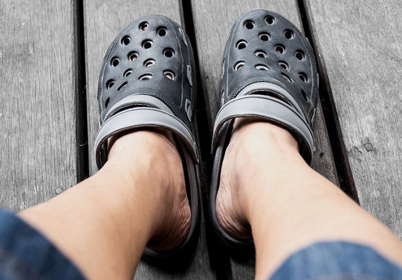
Prevention and smart self-check habits
You cannot prevent every case of foot melanoma, especially the types that are less clearly related to UV exposure. Still, smart habits help:
- check the tops and bottoms of your feet once a month
- look between the toes and around the heels
- inspect your toenails in good lighting
- use sunscreen on exposed feet when outdoors
- avoid tanning beds
- pay attention to one-nail changes that persist
- do not assume a nonpainful spot is harmless
A hand mirror, phone photo, or help from a partner can make foot checks easier. It is not glamorous, but neither is missing a cancer because it was hiding under a sandal strap.
Experience-based insights: what people often go through with foot melanoma
One of the most consistent themes in real-world foot melanoma stories is delay. Not because people are careless, but because the lesion often looks ordinary at first. Someone notices a dark patch on the sole and assumes it is dried blood. Another person spots a line on a toenail and blames old running shoes. Someone else thinks the area is a wart, corn, or fungus and tries over-the-counter treatment for months. By the time a biopsy happens, the lesion may be much thicker than it was when first noticed.
Another common experience is surprise. Patients often say they never expected melanoma on a foot because they associated melanoma with beach exposure, tanning, or a very obvious mole on sun-exposed skin. That false sense of “my feet are safe” can create a dangerous delay. It can be especially confusing when the lesion is not black or brown, because some foot melanomas are pink, red, or flesh-toned instead of dramatically pigmented.
People with toenail melanoma often describe a phase of misreading the signs. A dark band may be brushed off as a bruise. A podiatry or primary care visit may happen only after the streak widens, the nail becomes distorted, or dark pigment spreads onto nearby skin. For some, the turning point is simple: the mark does not grow out with the nail the way a typical injury should. That “wait, why is it still there?” moment matters.
After diagnosis, the next major experience is usually mental whiplash. What began as “I thought it was nothing” suddenly becomes pathology results, staging conversations, and treatment planning. Even when the melanoma is caught early, the emotional shift can be intense. Patients often describe fear, guilt about not coming sooner, and anxiety while waiting for biopsy margins or lymph node results. That emotional reaction is common and understandable.
Treatment experiences vary widely. Some patients have a small lesion removed and need close follow-up. Others need wider excision, reconstructive work, or treatment that affects walking for a while. Nail-unit surgery can be especially jarring because the foot is a daily-use body part, not a decoration. Recovery may involve wound care, activity limits, shoe adjustments, and frustration over how long “just a foot spot” can affect routine life.
For people with advanced disease, the experience often becomes more complex, involving oncology visits, scans, immunotherapy discussions, and long-term monitoring. Yet one encouraging pattern stands out: many patients say the most important lesson was learning to take small skin changes seriously, especially on areas they once ignored. In that sense, foot melanoma teaches a blunt but valuable lesson. The body’s quiet corners still deserve attention. If a spot is changing, persistent, or simply not behaving like a normal bruise or callus, getting it checked is not overreacting. It is good strategy.
Conclusion
Foot melanoma is uncommon compared with many other foot problems, but it is serious enough that missing it can have major consequences. The biggest danger is not always how dramatic it looks. The biggest danger is how ordinary it can seem at first.
If readers remember only a few things, let them be these: check the soles, check the toenails, do not ignore a changing spot, and do not assume a black streak is “just trauma” unless a qualified clinician says so. Early diagnosis gives doctors more options and patients a better chance at simpler treatment and a stronger outcome.